


 下载:
下载:
-
在固体与电解质溶液界面处,由于固体表面材料性质或(电极)溶液内电场作用,溶液中的带电离子运动或被吸附至固体表面及附近,在固体表面及其附近溶液界面形成厚度为几 nm至几 μm的固液界面层。固液界面广泛存在于纳米通道能量转换、锂电池和超级电容器、医学植入体以及微纳流器件等理论研究和应用中[1-5]。厚度为nm至μm的固液界面元素或离子(电荷)组成和动态变化与电化学反应、胶体结构、吸附作用和电容作用紧密相关,在上述应用场景中发挥着重要甚至决定性作用。计算机建模进行的仿真计算在固液界面的研究中被广泛地应用[6-9],模拟计算可以再现固液界面或者说液体表相双电层特定离子的交互变化趋势,但是无法得到结构和成分演化的信息。常规的实验测试分析手段,如透射电子显微镜(TEM)、原子力显微镜(AFM)可以提供详细的样品表面形貌变化的演化过程[10-11],却无法表征样品中化学成分信息;X射线光电子能谱(XPS)和X射线衍射(XRD)在固液界面分析中可以提供样品的物理和化学信息[12-13],但是较差的空间分辨率限制了其在固液界面分析中的应用。由于固液界面厚度在微纳尺度而测量探针易干扰甚至破坏界面结构,对固液界面结构进行无扰精细直接测量研究仍是当前固液界面研究中的一个技术难题。
离子束分析技术广泛应用在微束离子束分析中,因此MeV微束装置又被称为核微探针,微束开展离子束分析可以获得定量、高灵敏度和空间分辨的靶材料信息[14]。卢瑟福背散射分析技术(RBS-Rutherford Backscattering Spectroscopy)利用离子束与待测样品碰撞产生背散射能谱,通过对散射离子能量和计数进行统计分析就能获得靶原子深度与浓度信息的分布信息,是固体表面层元素成分、杂质含量和元素浓度的深度分布研究不可缺少的分析手段[15]。粒子激发X射线分析技术(PIXE-Particle Induced X-ray Emission)利用粒子轰击靶材,与靶材中的原子发生库伦相互作用电离产生特征X射线,通过探测这些特征X射线的能量和强度可以获得样品中的元素种类和含量[16]。这些离子束分析方法具有无损且元素分析灵敏度高的显著优点,结合微束成像技术还可以得到元素微区分布甚至是三维分布信息,但是传统上只能直接用于固体薄样品的元素组分与深度分布的分析。为了拓展离子束分析技术的使用范围,2018年Saito等[17]利用外束卢瑟福背散射分析方法,通过使用300 nm氮化硅和150 nm金复合薄膜的真空窗,在大气环境中观察到了硝酸银溶液中银离子沉积在金电极表面的现象。但是从研究结果来看,高深度分辨、空间分辨和元素分析精度的离子束分析需要超薄窗样品,并在高真空且低环境本底中进行。为了解决真空中分析固液界面的困难,1987年Forster等[18]首先将离子束透射1 μm厚的氮化硅薄窗后,基于卢瑟福背散射技术分析电极表面的元素分布来研究电化学过程,并利用弹性背散射离子能谱获得了电极表面的元素组份信息。2009年Hightower等[19]采用150 nm厚的氮化硅薄窗进行卢瑟福背散射实验来分析电化学电极界面的元素分布。但是这些早期利用卢瑟福背散射进行固液界面的离子束分析工作由于采用的分析窗口过厚,限制了固液界面元素深度结构解析精度[17-19]。2011年Yang等[20]利用聚焦离子束和二次离子质谱方法,通过聚焦离子束凿出微米小窗暴露液体界面的方法在线分析了电极界面处所暴露的液体表面的元素分布,这种技术破坏了界面结构,影响了界面元素的结构和分布。
随着薄膜制备技术的发展,采用更薄的纳米膜用于制备真空窗或电极成为可能,从而可大大提高离子束分析的元素纵向分辨。为了实现真空环境下对固液界面的元素高精度结构分析,我们制备了可以稳定运用于离子束分析的固液界面电极探针,利用PIXE和RBS技术获得了固液界面的元素信息及其随电场的变化动态过程,还通过PIXE微束成像技术获取了固液界面特定离子的交互变化成像图。
-
基于离子束分析技术的原理和固液界面的分析要求,设计了如图1(a)所示的固液界面电极探针,探针系统用薄膜将溶液封装在一个腔体内,纳米薄膜既是离子束入射的真空窗又能实现对溶液的封装。作为隔离真空和液体的密封窗,纳米薄窗应满足致密性良好、力学强度优异、透光率高及理化性能稳定这几个基本要求,并能承受离子束分析时的辐照损伤。经过多种尺寸和厚度薄窗样品的气压强度测试实验,选用了基于200 μm厚的硅作为支撑框架,中间部分是尺寸为100 μm×100 μm、厚度为15 nm的低压气相沉积生长的氮化硅薄膜作为纳米薄窗,硅框架与氮化硅薄膜中间窗口呈54°。作为固液界面电极探针的工作电极,应该兼具薄且导电性优异的特性,采用铝作为电极可以避免在卢瑟福背散射分析中对水溶液中重元素的干扰。本文采用真空蒸发镀膜的方法在氮化硅薄窗上制备了实际成膜厚度为10.4 nm的铝电极作为固液界面电极探针的电极。图1(b)是使用原子力显微镜观测的铝电极表面形貌示意图,可以看出,铝电极表面均匀性较好,划痕法测得的铝膜厚度为10.5 nm左右,与高精度天平称重法测量结果(10.4 nm)基本相符。
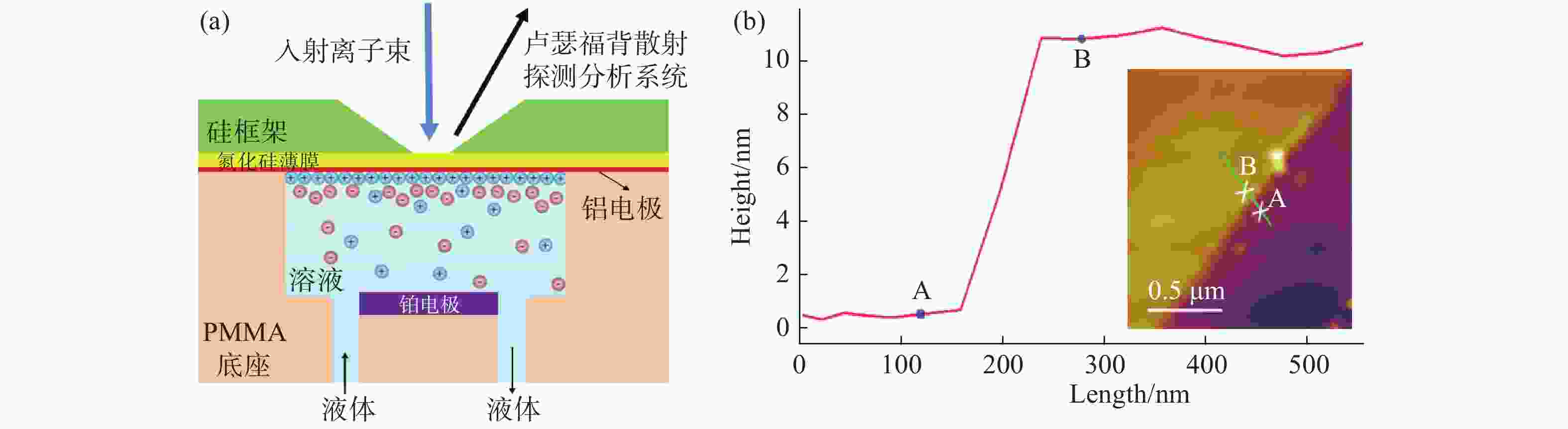
图 1 (在线彩图) 固液界面电极探针设计示意图(a)及使用原子力显微镜观测的铝电极表面,以及划痕法测量的电极厚度示意图(b)
固体液体界面探针的构成如下:在聚甲基丙烯酸甲酯(PMMA-polymethylmethacrylate,俗称为有机玻璃)底座上制备出体积为3 mm×3 mm×3.5 mm溶液存储腔,有机玻璃腔体透明度高,便于观测,确保注入溶液过程中没有产生气泡且溶液充分填满整个腔体;使用硅作为支撑框架薄膜厚度为15 nm,尺寸是100 μm×100 μm的氮化硅薄膜作为真空窗来进行隔离液体腔和真空分析环境,并用环氧树脂胶将硅框架部分密封在有机玻璃底座上。氮化硅薄膜面向液体腔层真空蒸发镀有10 nm铝作为工作电极,采用铂电极放置在液体腔底部作为辅助电极,这样固液界面电极分析系统可通过实时对电极外加偏压实现在真空环境中对固液界面元素和电化学过程的动态分析。实验待测溶液通过化学惰性好的聚醚醚酮(PEEK-polyetheretherketone)管链接的PEEK阀门输入到液体存储槽内,并使用环氧树脂作为底座和管道以及阀门之间的密封材料,灌入不同的溶液即可构建不同的固液界面实验系统。探针整体放入离子束分析靶室测试,维持真空度为10–6 Pa,可以满足实验真空度需求。
-
实验所用的溶液是氯化钡和氯化镧溶液。因为卢瑟福背散射能谱的产额对于原子序数非常敏感,对于轻基体元素上的重元素有较强的灵敏度,所以选用Ba元素和La元素作为溶液中的特征元素进行数据收集能直观地观测到变化趋势,不会受到与轻元素能谱产生重叠等影响。使用固液界面电极探针时,如果液体发生渗漏将会引起靶室内真空度下降,轻则引起设备自动断电停止工作,重则会导致真空泵以及靶室内探测器的损坏,所以实验中放入的每一个固液界面探针都提前进行了真空环境24 h漏液测试。
本文的离子束分析实验是利用复旦大学现代物理研究所加速器核微探针系统完成的。该微束系统使用Si(Au)面垒探测器记录背散射粒子,放置在与束流呈165°的方位,对于3 MeV的质子其能量分辨率可达15 keV,有效面积是28 mm2;粒子激发X射线的Si (Li)探测器灵敏面积为80 mm2,Be窗厚度为12 μm,对于5.9 keV的X射线其能量分辨率为150 eV,放置在与束流呈135°的位置。
-
为了获得固液界面的结构信息及在电场作用下的界面组成的演化过程,本文使用束斑面积是68.36 μm×93.75 μm 的1.0 MeV质子束,以及束斑面积是68.36 μm×93.75 μm 的2.0 MeV的氦离子束进行离子束分析实验。首先采用粒子激发X射线技术对制备的固液界面探针进行了元素微区分析,之后结合卢瑟福背散射技术对固液界面进行了元素分析。
-
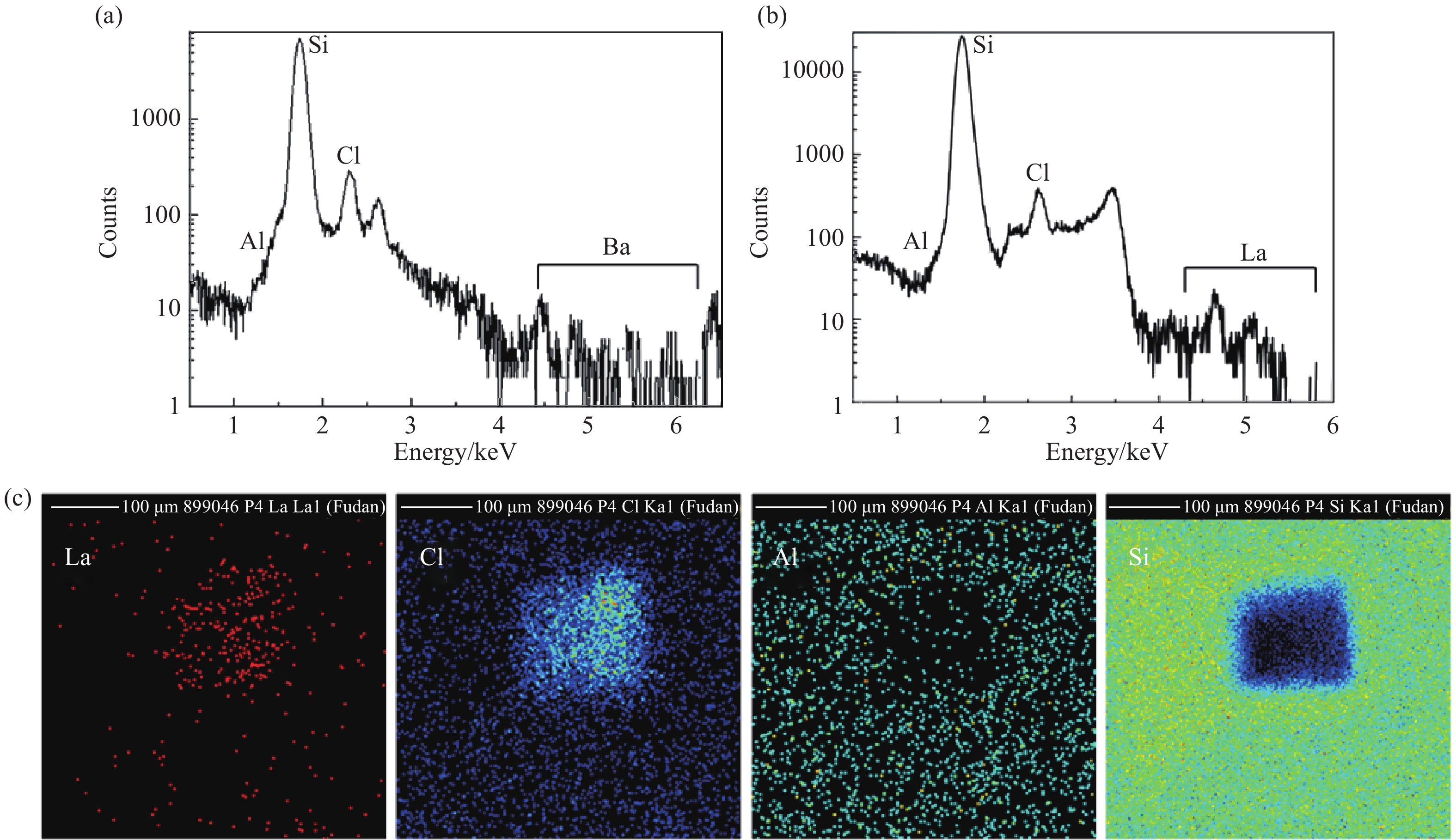
当利用PIXE进行样品分析时,分析离子束在材料中的射程决定了所获得的元素来源。利用复旦大学核微探针系统,通过使用2.0 MeV的He+离子束和1.0 MeV质子束分别分析了距固液界面11和26 μm深度内溶液中的元素分布情况。图2(a)使用1.0 MeV的质子作为入射离子得到了0.01 mol/L的BaCl2溶液PIXE能谱(扫描范围500 μm×500 μm,测量1 h),可以看出,由于低本底溶液浓度的影响,Ba元素的计数率很低。图2(b, c)为固液探针内注入1 mol/L的LaCl3溶液后,获取的分析区的 PIXE能谱和对应的元素二维成像分布图(扫描范围500 μm×500 μm,测量5 min)。从图中可以看出,基于我们设计的固液探针系统,在真空环境下通过PIXE分析可以清晰获取溶液内的元素二维分布。La元素和Cl元素仅分布在100 μm×100 μm的固液界面薄窗区域,硅元素主要分布在薄窗的硅框架。铝元素由于电极太薄而探测计数少,基本淹没在硅元素特征峰内,无法清晰成像。PIXE测量只能给出入射离子射程内的元素总量,无法给出这些元素在固液界面附近液体内的深度分布。
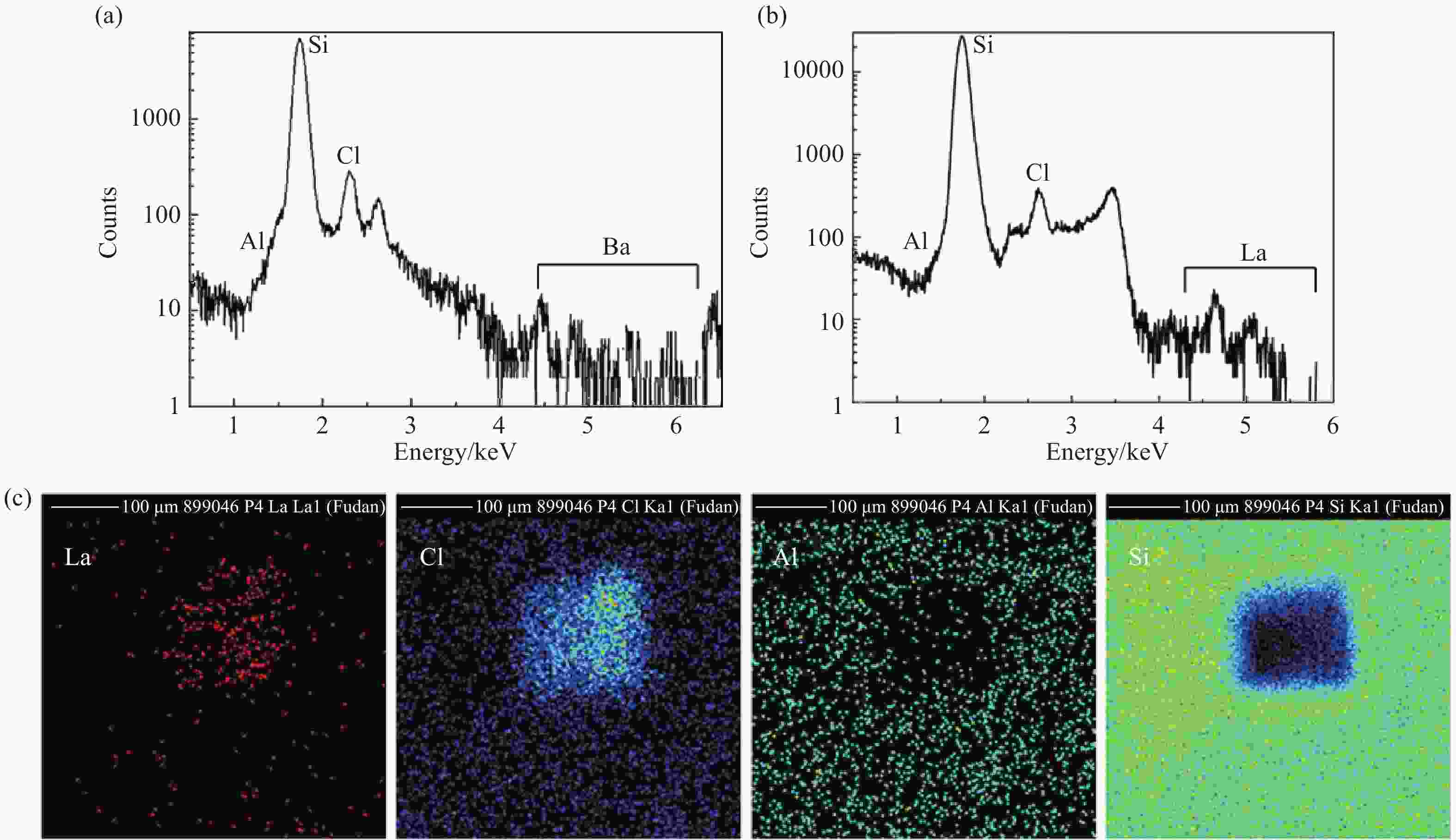
图 2 (在线彩图) 利用固液界面探针测量得到的PIXE能谱
-
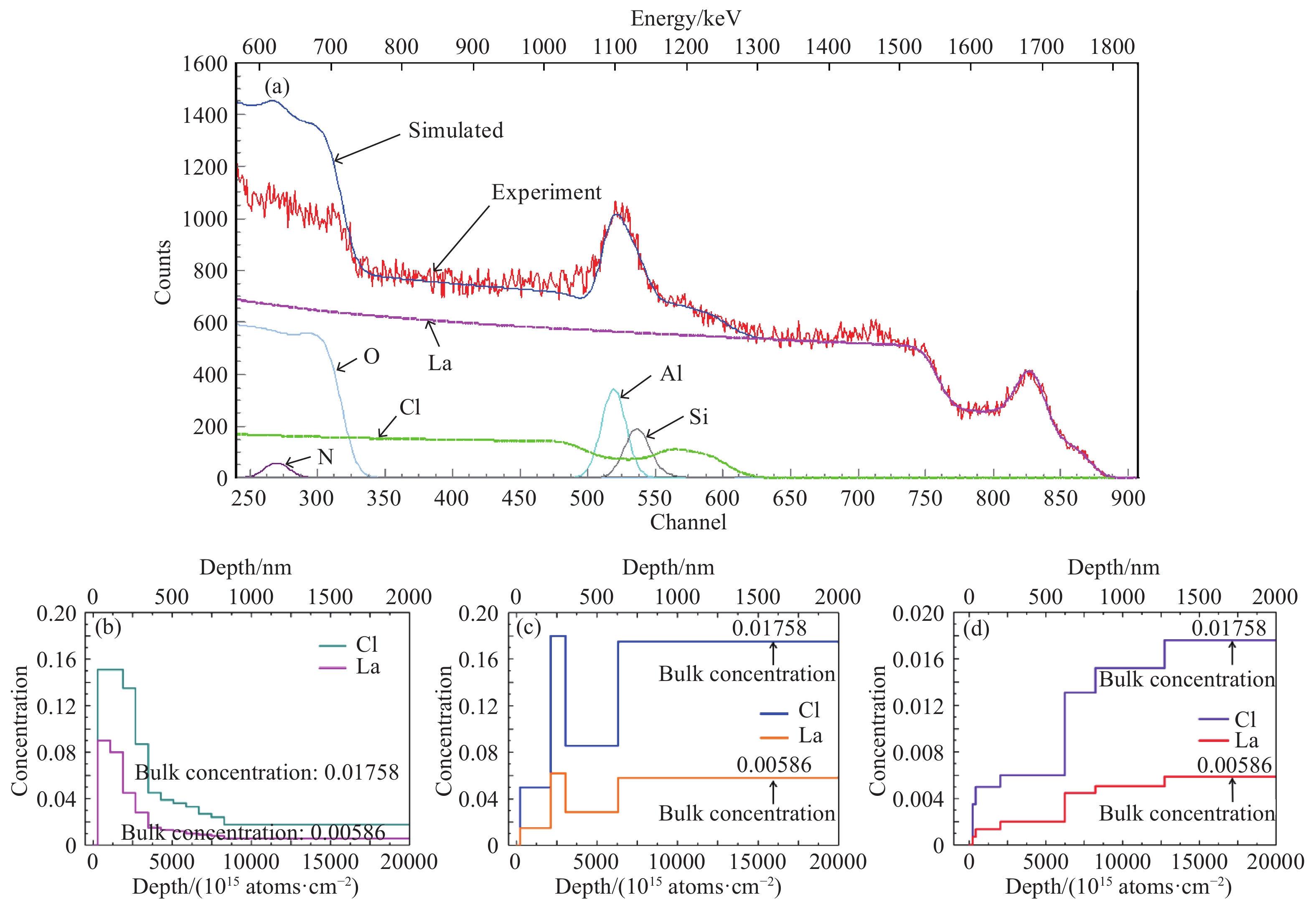
卢瑟福背散射能谱分析可开展材料表面高精度的元素深度分析,利用粒子激发X射线微束分析获得各种元素的二维分布后[图2(c)],选定固液界面探针的薄窗区域,开展微束低分析截面的RBS测量。为了在线制备不同的固液界面环境,对固液界面探针的铝电极先后加偏压–2.3,0,+2.3 V,都进行了5 h的RBS能谱采集测量。图3(a)为测量得到的固液探针腔体充满1 mol/L LaCl3溶液时的RBS分析能谱(0 V)和使用SIMNRA[21]软件解谱得到的元素浓度随深度的分布。
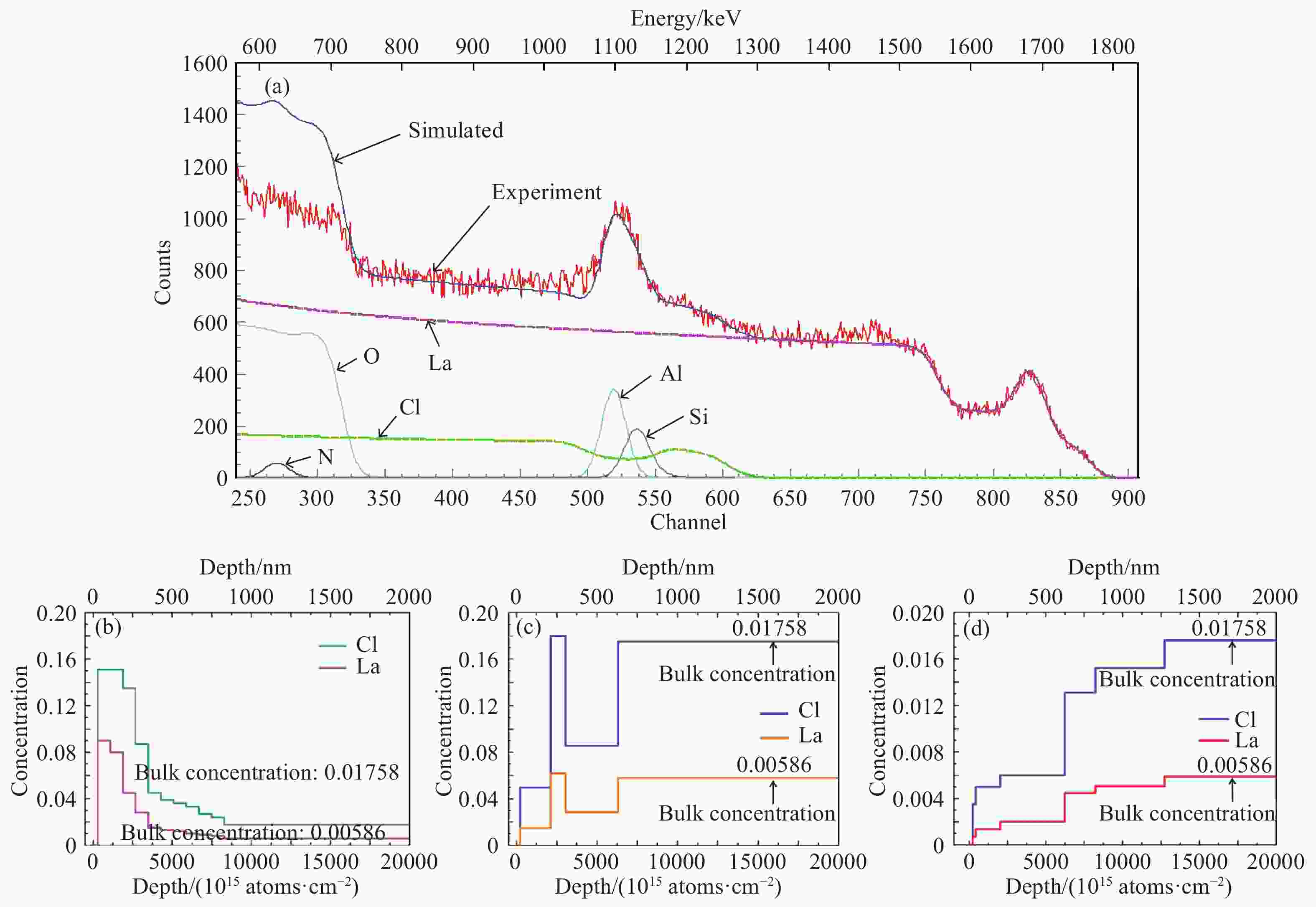
图 3 (在线彩图) 固液界面电极探针(1 M氯化镧溶液)的RBS实验能谱(偏压0 V,2.0 MeV He+,5 h采集)和各元素SIMNRA计算解析,解谱拟合得到的偏压–2.3 V电极界面附近Cl和La元素在溶液中的浓度随深度的分布
从图3(a)可以看出,固液界面电极探针的RBS能谱图中背散射He+离子的La,Cl,Si,Al,O,N元素能谱峰和能谱台阶明显,SIMNRA拟合总谱La,Cl,Si,Al元素与实验数据基本吻合,O元素的拟合谱与实验数据有一定的差异,这是因为溶液在长时间的辐照下可能产生了辐射分解[22],在这个过程中产生了相应的化学反应导致O元素被消耗。图3(b~d)是解谱拟合得到的同一样品在–2.3,0,+2.3 V偏压时Cl元素和La元素浓度随深度的分布示意图,可以看出,在铝电极表面阴阳离子浓度与溶液配制体浓度(bulk concentration)有一定的区别。在负压铝电极表面,LaCl3溶液中Cl元素浓度约9倍于体浓度,La元素浓度约15倍于体浓度,溶液近表面270 nm以内,Cl元素相对浓度与La元素相对浓度之比小于它们在本底溶液中的配比3:1,说明在这个范围以内La元素相对含量比Cl元素高,即La元素在电极表面附近聚集,此后至820 nm深度Cl元素和La元素的浓度逐渐递减至配制体浓度。在无偏压时,电极表面溶液中Cl元素和La元素的浓度先呈增加趋势,在300~600 nm处有剧烈的下降,距离电极表面600 nm后溶液Cl元素和La元素的浓度与配制浓度相符。300~600 nm这段Cl元素和La元素的浓度下降可能是由于分析区域在离子束分析过程中由于辐照产生的微气泡造成。当改变电极偏压至+2.3 V时,电极附近溶液由于浓度差异分为六层,在铝电极表面180 nm以内,Cl元素与La元素相对浓度之比大于两者的本底浓度之比3:1,说明Cl元素被吸引到近表面,此后至1 250 nm两者浓度之比呈逐级升高趋势,最终达到了溶液的配制体浓度,还可以得出本次实验中获得的液体样品的最优深度分辨率为20 nm,对应着溶液中的第一层。
对比固液界面电极施加偏压前后的元素深度分布可以得出,在金属电极固液界面处,电解液中的阴阳离子受电场驱动的非平衡分布达到了微米尺度。这种离子分布形态与由于固体表面带电引起的几纳米至几十纳米范围的双电层离子分布不同[23],更接近于电化学电极表面的化学反应过程中微米范围的溶液扩散层部分元素的分布[24]。基于我们制备的固液界面电极探针,利用卢瑟福背散射分析技术成功得到了固体电极与电解液界面的元素分布。从卢瑟福背散射能谱解谱得到的固液界面的元素随着深度分布的示意图中可以看出,实验获取了溶液中扩散层部分浓度变化情况且获取的深度分辨率最优为20 nm范围,对固液界面铝电极外加负压时La元素富集在固液界面附近,对固液界面铝电极外加正压时Cl元素富集在固液界面附近。在He+离子束RBS分析实验前后,我们对固液界面探针的薄窗进行了光学显微镜观察对比,充满液体的固液界面探针薄窗在约1.0×10¹⁸ ions/cm2的He+离子辐照(15 h)前后没有显著变化,液体没有渗漏,表明其抗辐照性能良好。
-
本文介绍了一种可以用于真空环境离子束分析的固液界面探针设计,该探针的纳米薄窗能够承受1个大气压强差和高剂量的分析离子束辐照损伤。利用该探针成功实现了真空环境下的固体液体界面的原位元素分析,初步获取了固液界面微米尺度内的构成信息。利用微束PIXE分析和RBS分析获取了不同实验条件下固液界面元素组分信息,通过PIXE成像技术获得了固液界面元素的二维分布信息;利用RBS分析获取了电极电解液界面附近元素的几十nm至μm的深度分布信息,以及外加电压的调控下微米尺寸扩散层范围内溶液中特征离子相对浓度的变化关系。该项工作表明,高真空环境的离子束分析与成像技术可以开展固液界面和液体中的元素成分和结构演化的形态信息的相关研究,这将利于传统离子束技术在电化学、能源材料、界面科学等领域开展更多的应用。未来对固液界面探针进行优化改进,并结合高分辨卢瑟福背散射分析技术和高分辨微束技术,可以进一步提高固液界面元素分析的空间分布分辨率并深入探究外加电场的调控下固液界面的微观反应机理。
-
摘要: 原位实时地高精度测量固液界面的元素或离子(电荷)组成和动态变化对于界面反应和相互作用研究非常重要,但是传统的高分辨离子束分析实验在真空环境中不能直接测量液体样品。本文研制了一种固体-液体界面探针,该探针使用氮化硅-铝纳米复合膜作为真空密封窗和电化学电极,利用复旦大学核微探针成功开展了真空中固体-液体界面探针0.01 mol/L氯化钡和1 mol/L氯化镧溶液样品固体-液体界面的卢瑟福背散射(RBS)分析和粒子激发X射线(PIXE)分析。实验结果表明,真空环境下,固液界面探针纳米薄窗可承受2 MeV He+离子注量为1.0×10¹⁸ ions/cm2的辐照。微区PIXE分析成功获得了固液界面探针结构的元素分布。通过对卢瑟福背散射能谱进行分析,获取了20 nm分辨的电极界面微米深溶液中的La, Cl元素浓度。在1 mol/L的LaCl3固液界面电极表面,负电压(–2.3 V)时电解质离子在电极表面高浓度聚集,正电压(+2.3 V)时电解质在电极表面呈低浓度分布,在约1 250 nm深处电解质溶液趋向于体浓度。Abstract: The in-situ and real-time high-precision measurement of the composition and dynamic change of elements or ions (charges) at the solid-liquid interface with nano-to-micron thickness is very important in the understanding of the interface interaction and reaction, while traditional high-resolution ion beam analysis can not directly measure liquid samples in vacuum environment. In this paper, a solid-liquid interface probe in vacuum was developed. The probe used Si3N4-Al nanocomposite membrane as vacuum sealing window and electrochemical electrode. The Rutherford Backscattering Spectroscopy (RBS) analysis and particle-induced X-ray Emission (PIXE) analysis with the solid-liquid interface probes of 0.01 mol/L BaCl2 and 1 mol/L LaCl3 solution were successfully carried out using Fudan University nuclear microprobe. The experimental results show that the nano-window of solid-liquid interface probe can withstand the irradiation of 2 MeV He+ ions with a dose of 1.0×10¹⁸ ions/cm2 in vacuum. The distribution of structure elements in solid-liquid interface probes was successfully obtained by PIXE analysis. The concentration of La and Cl in micron deep solution of electrode interface was obtained by Rutherford backscattering analysis with 20-nm-resolution. On the surface of 1 mol/L LaCl3 solid-liquid interface electrode, electrolyte ions accumulated at a high concentration at negative voltage (–2.3 V), while electrolyte ions distributed at a low concentration at positive voltage (+2.3 V), and electrolyte solutions tended to bulk concentration at a depth of about 1 250 nm.
-
图 2 (在线彩图) 利用固液界面探针测量得到的PIXE能谱
(a) 1 MeV质子,1 h采集,0.01 mol/L BaCl2溶液;(b) 2 MeV He+,5 min采集,1 mol/L LaCl3溶液;(c) 2 MeV He+, 5 min采集,1 mol/L LaCl3溶液元素微区分布成像。
-
[1] VANDER H, FRANK H J, BONTHUIS D J, et al. Nano Letters, 2007, 7(4): 1022. doi: 10.1021/nl070194h [2] BIEKER G, WINTER M, BIEKER P. Chemical Physics, 2015, 17(14): 8670. doi: 10.1039/C4CP05865H [3] DU X, GUO P, SONG H H, et al. Electrochimica Acta, 2010, 55(16): 4812. doi: 10.1016/j.electacta.2010.03.047 [4] KIM S M, BURNS M A, HASSELBRINKE F. Analytical Chemistry, 2006, 78(14): 4779. doi: 10.1021/ac060031y [5] BOCQUET L, CHARLAIX E. Chemical Society Reviews, 2010, 39(3): 1073-0. doi: 10.1039/b909366b [6] CHEIN R, CHEN H, LIAO C. Journal of Electroanalytical Chemistry, 2009, 630(1-2): 1. doi: 10.1016/j.jelechem.2009.01.025 [7] KANG S, SUH Y K. Microfluid Nanofluid, 2009, 6(4): 461. doi: 10.1007/s10404-008-0321-5 [8] KISLENKO S A, SAMOYLO I S, AMIROV R H. Physical Chemistry Chemical Physics, 2009, 11(27): 5584. doi: 10.1039/B823189C [9] DEYOUNG A D, PARK S W, DHUMAL N R, et al. Journal of Physical Chemistry C, 2014, 118(32): 18472. doi: 10.1021/jp5072583 [10] MEHDI B L, QIAN J, NASYBULIN E, et al. Nano Letters, 2015, 15(3): 2168. doi: 10.1021/acs.nanolett.5b00175 [11] SIRETANU I, EBELING D, ANDERSSON M P, et al. D Scientific Reports, 2014, 4(1): 4956. doi: 10.1038/srep04956 [12] OSWALD S, NIKOLOWSK K, EHRENBERG H. Analytical & Bioanalytical Chemistry, 2009, 393(8): 1871. doi: 10.1007/s00216-008-2520-z [13] LIU L J, CHEN L Q, HUANG X J, et al. Journal of The Electrochemical Society, 2004, 151(9): A1344. doi: 10.1149/1.1772781 [14] 杜广华. 原子核物理评论, 2012, 29(4): 371. doi: 10.11804/NuclPhysRev.29.04.371 DU Guanghua. Nuclear Physics Review, 2012, 29(4): 371. (in Chinese) doi: 10.11804/NuclPhysRev.29.04.371 [15] 杨福家, 赵国庆. 离子束分析[M]. 上海: 复旦大学出版社, 1985. YANG Fujia, ZHAO Guoqing. Ion Beam Analysis[M]. Shanghai: Fudan University Press, 1985. (in Chinese) [16] 任炽刚. 质子X荧光分析和质子显微镜[M]. 北京: 原子能出版社, 1981. REN Zhigang. Particle Induced X-Ray Emission and Protom Scanning Microscope[M]. Beijing: Atomic Energy Press, 1981. (in Chinese) [17] SAITO M, HOLM K, BREGOLIN F L, et al. Surface and Interface Analysis, 2018, 50: 1149. doi: 10.1002/sia.6396 [18] FORSTER J S, PHILLIPS D, GULENS J, et al. Nucl Instr and Meth, 1987, 28(3): 385. doi: 10.1016/0168-583X(87)90180-7 [19] HIGHTOWER A, KOEL B, FELTER T. Electrochimica Acta, 2009, 54(6): 1777. doi: 10.1016/j.electacta.2008.10.027 [20] YANG L, YU X Y, ZHU Z H, et al. Lab on a Chip, 2011, 11(15): 2481. doi: 10.1039/c0lc00676a [21] MAYER M. SIMNR A. A Simulation Program for the Analysis of NRA, RBS and ERDA[C]// DUGGAN J L, MORGAN I L.AIP Conference Proceedings of the Fifteenth International Conference on the Application of Accelerators in Research and Industry. New York: American Institute of Physics, 1999: 541. [22] BOUQUILLON A, DRAN C, LAGARD G, et al. Nucl Instr and Meth B, 2002, 188(1-4): 156. doi: 10.1016/s0168-583x(01)01066-7 [23] PAZGARCIA J M, JOHANNESSON B, OTTOSEN L M, et al. Electrochimica Acta, 2014, 150: 263. doi: 10.1016/j.electacta.2014.10.056 [24] GRAHAME D C. Chemical Reviews, 1947, 41(3): 441. doi: 10.1021/cr60130a002 -

 点击查看大图

图(3)
计量
- 文章访问数: 1421
- HTML全文浏览量: 453
- PDF下载量: 45
- 被引次数: 0
 甘公网安备 62010202000723号